Technologies
- Bone replacement from own blood (PRF Technology)
- Dental CT
- Dental microscope
- Dental laser
- Intraosseous anaesthesia
- Digital implantation

Bone replacement from own blood (PRF Technology)
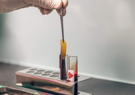
A few decades ago, people realized that their own blood helps the human body to heal optimally. The essence of this revolutionary new technology is that bone and tissue replacements are performed with plasma made from the patient’s own blood instead of synthetic bone replacements. Compared to traditional bone substitutes, this “own material” is more beneficial because it is full of agents that stimulate regeneration, e.g. with growth factors, fibrin, white blood cells, which are impermeable to bacteria.
Procedure:
- Laboratory tests before surgery: laboratory tests for certain blood parameters are recommended 1-2 weeks before surgery.
- Blood sampling: Immediately before the operation, depending on the size of the intervention, we take 2-8 tubes of blood from the patient.
- Centrifugation of the collected blood: the blood is centrifuged in a special device. As a result, its cellular elements separate into red blood cells and white blood cells. We use the latter for the surgery.
- The gelatinous substance obtained in this way is implanted in the surgical area by itself or mixed with a bone substitute.
Advantages of the procedure: The heaing is almost half comared to the tradicional methods.
- In most cases, the healing is almost half compared to the traditional methods.
- Thanks to the faster recovery, the feeling of well-being after surgery is also significantly better.
- From a material point of view, since the method has at least halved the price of previous larger bone replacements, patients can access these interventions more cheaply.
- There is no risk of rejection: it is not a foreign bone substitute that enters the body, but the material produced by the patient’s body itself.
- After tooth extraction, inflammation can be avoided by placing it in place of the tooth extraction.
- The original size and size of the jawbone can be maintained, because the surrounding tissues suffer significantly less degeneration, the blood vessels and nerves weave through the area many times faster, giving the opportunity for easy insertion of the future implant (socket preservation).
We recommend it for all surgeries!
Contraindication:
- taking stronger anticoagulants;
- malignant hematopoietic disease.
Press releases
GGDent’s own CT scanner for dental surgeries
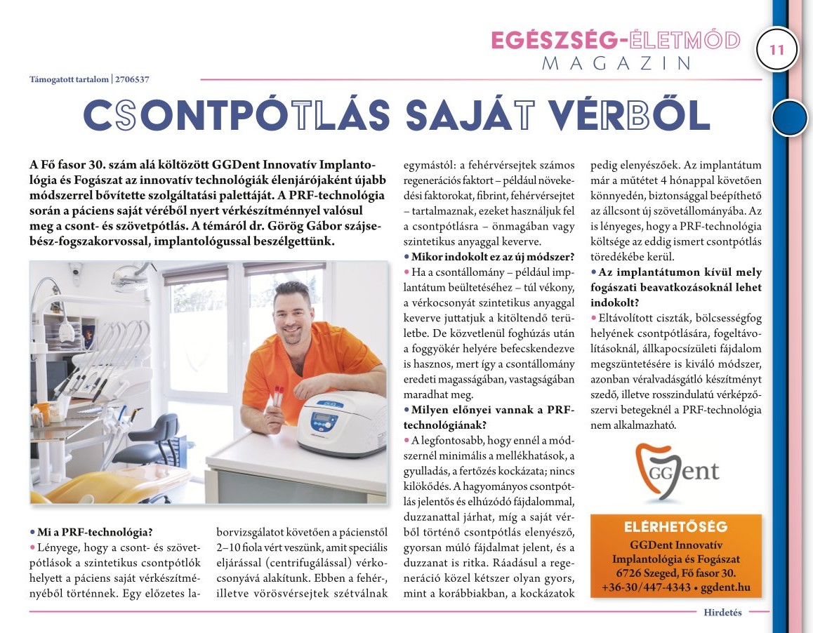
GGDent Innovative Implantology and Dentistry, which has moved to 30 Fő fasor, has expanded its range of services with a new method as a leader in innovative technologies. During the PRF technology, bone and tissue replacement is carried out with a blood product obtained from the patient’s own blood. On the topic, we talked with Dr. Gábor Görög, oral surgeon dental specialist and implantologist.
Délmagyarország Health magazine annex 2024.03.30.
Dental CT Scan
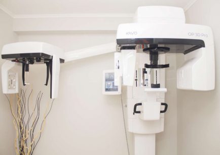
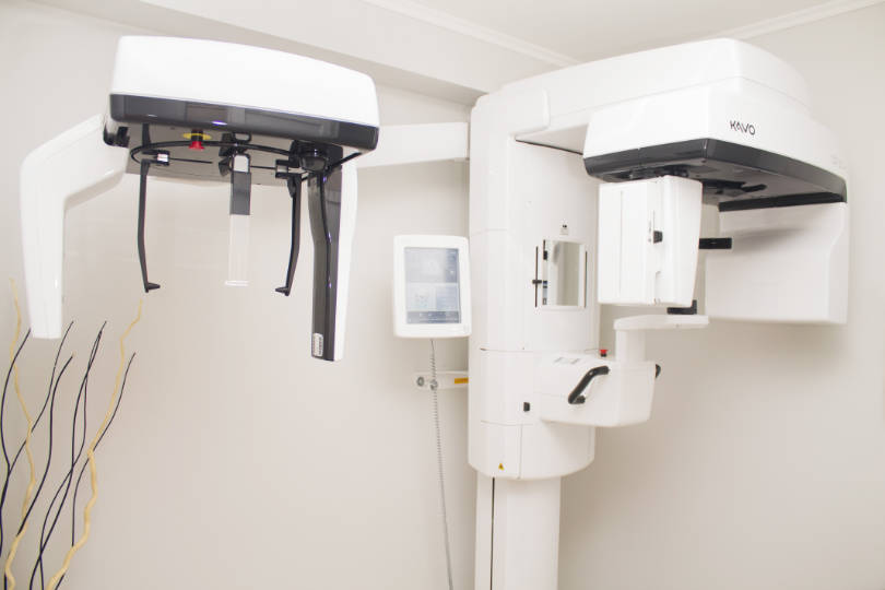
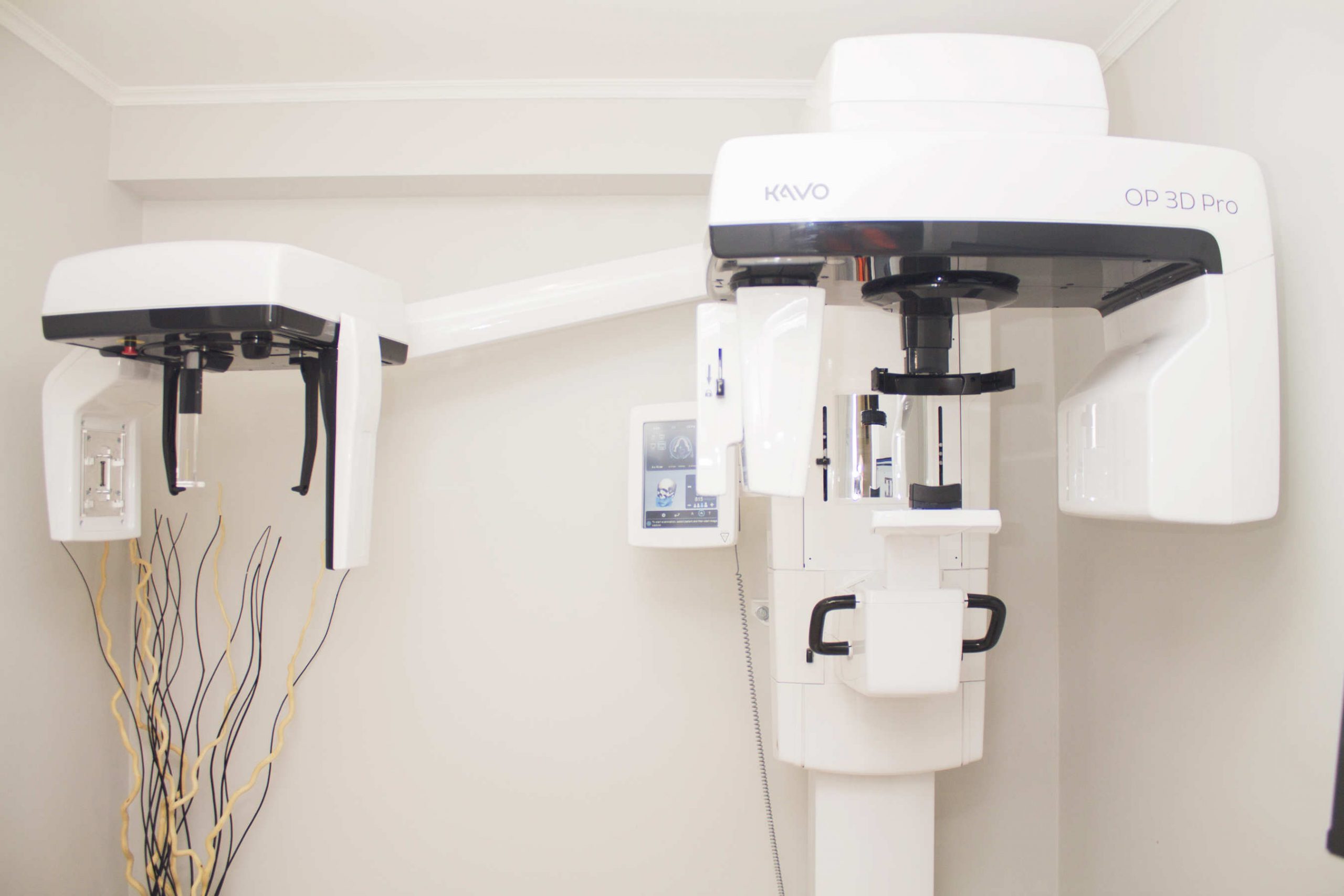
In dentistry, only panoramic X-rays were previously available, which only allowed the teeth and bone structure to be seen in 2 dimensions. However, in our practice, we have a Cone Beam CT (CBCT) imaging device, which allows us to obtain a three-dimensional image of the state of the teeth, avoiding the distortions and uncertainties that can occur on traditional X-rays. The type of equipment we use is a KaVo OP 3D Pro Ceph. The device rotates around the patient’s head in 10-14 seconds and in 2-3 minutes the image is taken and given to the patient on CD.
The CBCT is a great advantage in implantology, oral surgery and root canal treatment, but also in a simple general inspection or in the diagnosis of tooth pain, because we know exactly where the intervention is needed, so that the intervention is only exactly as big as absolutely necessary. It is most widely used in implantology, where the position of not only the bones but also soft tissues can be measured with millimeter accuracy. It is possible to determine precisely where bone replacement is needed, or the position of the sinus cavity and its inflammation and the position of the nerves in the lower jaw. In oral surgery, it is of particular importance regarding the removal of wisdom teeth. It shows the position of the root of the tooth in relation to the larger nerve in the jaw, so that possible damage can be avoided. In root canal treatment, CBCT can also be used to show the site and any collateral canals, fractures and possible inflammation.
3D CBCT scans at GGDent Dental and Implant Center:
- CBCT full dental arch
- CBCT sinus
- panoramic X-ray
- lateral X-ray
- TMI right side CBCT
- TMI left side CBCT
- CT scan of 1-3 teeth
- MANDIBULA/MAXILLA CBCT
Press releases

GGDent’s own CT scanner for dental surgeries
At the end of the year, the GGDent Dental and Implantation Centre in Szeged replaced its panoramic X-ray machine with a CT scanner. The device, which is most useful for a better and safer implantation of dental implants, provides a 3D image of the entire facial skull.

Dental microscope
About dental microscope
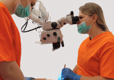
Dental microscope is a high-tech device that allows the doctor to see the area to be treated at a magnification of up to 30-40 times, thus enabling more precise diagnosis and treatment. With its use, we can establish a much more accurate diagnosis, thereby increasing the effectiveness of the treatment.
In what cases can it be used?
Dental microscope is most often used for root canal treatment, thanks to which the success rate is significantly higher compared to traditional root canal treatment. It creates a very detailed picture of the dental structures, and with its built-in high-brightness light, even hard-to-see areas become clearly visible.
In addition to root canal treatment, it can be used in many cases, such as opening invisible cracks, repairing perforations, treating gum diseases, etc.
Why is microscopic root canal treatment preferable?
- more precise diagnosis thanks to the high-resolution image;
- cracks and mini-decays that are difficult to see with the naked eye can be noticed immediately;
- much higher success rate compared to traditional root canal treatment;
- hidden root canals can also be revealed;
- less healthy tooth material needs to be removed;
- there is a greater chance of saving the tooth;
- safer, faster, more efficient treatment.
How does a microscopic root canal treatment take place?
- Local anesthesia.
- Placing rubber dam for perfect isolation (to avoid possible infections).
- Small drilling in the crown of the tooth to access the root canal.
- Measuring the length of root canals.
- Cleaning of root canals.
- Draining of channels.
- Insertion of root filling material.
Our personal consultation is free, please contact us for more information: +36-30-447-43-43.
Dental laser
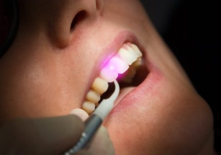
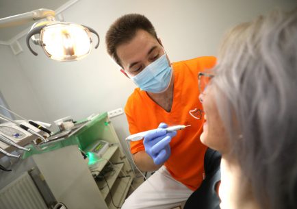
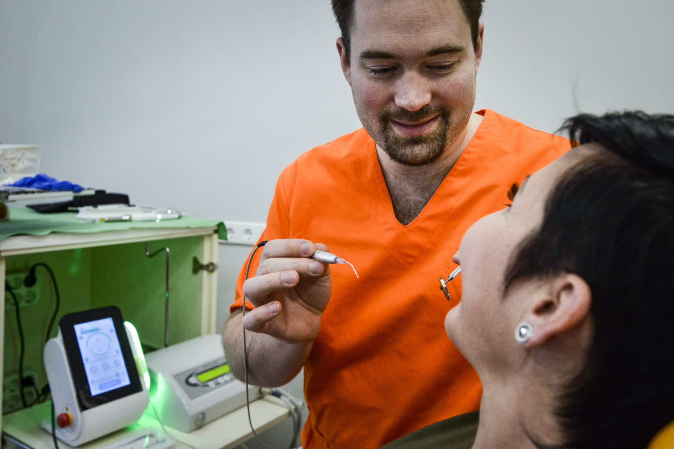
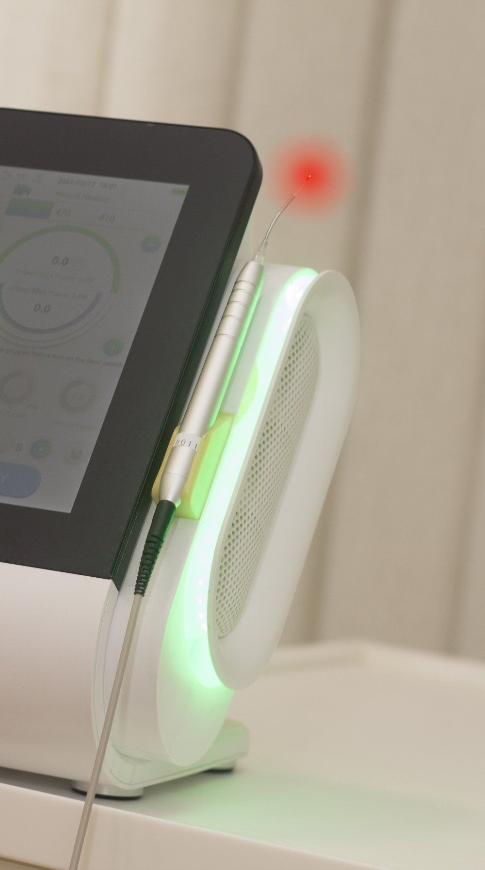
Previously used mainly in industrial applications, lasers have in the last 25 to 30 years also entered the field of dentistry, offering many advantages over traditional dental treatments. The red light from the combined device used in these procedures is used to treat problems on the surface of the mucous membrane, while the infrared light is used to treat deeper areas. However, it is important to stress that laser treatment is in most cases a supplementary treatment next to conventional methods.
Its main advantages are:
- anti-inflammatory: in most cases, inflammation is reduced by half compared to previous methods, thanks to the biostimulation effect produced by the laser
- haemostatic (reduces the bleeding): it has its significance in oral surgery, especially for patients with haemophilia
- pain reduction: pain after procedures is reduced to a minimum
- faster wound healing, regenerative effect: healing time is almost halved. This is a major advantage for the elderly and diabetics.
- the laser kills bacteria: excellent results for root canal treatment
- acceleration effect: for example, in the case of teeth whitening, it reduces the time needed for the procedure, which would otherwise take 1-1.5 hours, to a fraction of the time by enhancing the effect of the whitening material
- regeneration in the treatment of osteoporosis
- helping the healing of mucosal lesions caused by aphthae and viruses
It can be used in almost the entire range of dental procedures, including:
- in oral surgery: for cutting soft tissues
- implantation
- root canal treatment
- removal of dental neck sensitivity
- treatment of herpes, aphthae
- bone grafting
- teeth whitening
- treatment of periodontal diseases
- gum surgery
- treatment of dry mouth
- removal of dental caries
Intraosseous anaesthesia
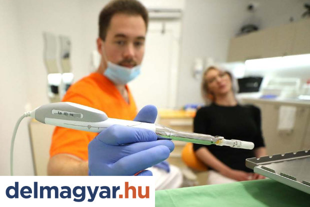
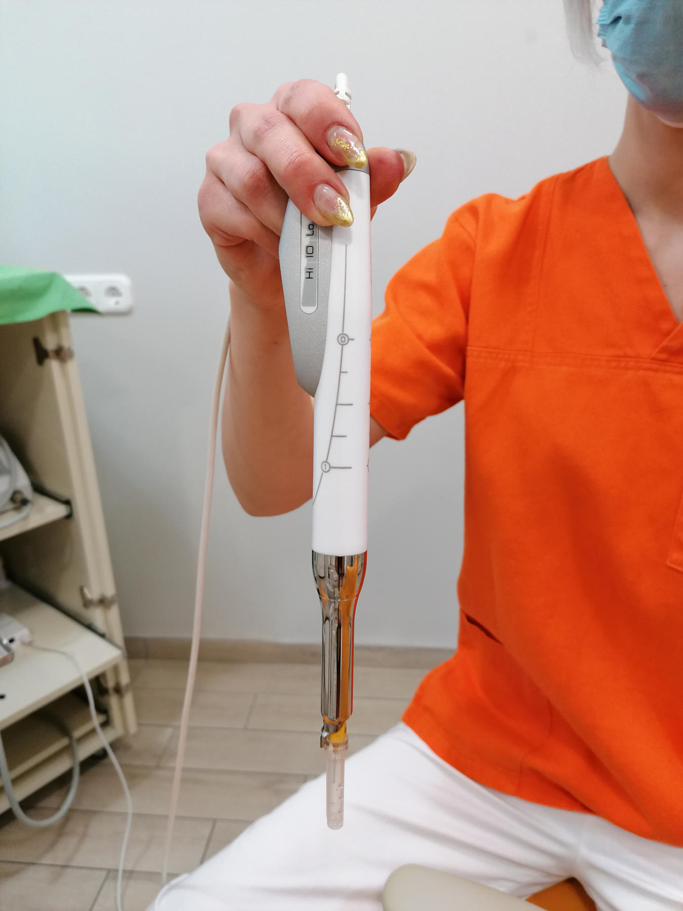
Intra-osseal anaesthesia is a novelty in dentistry, the result of a rethinking of an older technology. The machine we work with is called the Quicksleeper. The idea is that for a planned procedure, such as a filling or a root canal, the patient first needs just a few drops of anaesthetic to be injected into the gum between the two teeth, which makes the area suitable for the anaesthetic to be administered. Then, using a specially designed machine and a special needle, we can painlessly inject the anaesthetic fluid directly into the nerve endings at the root of the tooth. This actually numbs some of the teeth close to the injection site without numbing most of the gums or face until the anaesthetic takes effect. For the injection, a panoramic X-ray is always needed to know how close the roots are to each other and whether the thin needle can fit next to them.
Main advantages:
- numbness is almost immediate, no waiting time to start the work
- the patient has no speech difficulties after the procedure
- the anaesthesia is successful 99.9% of the time, so there is no need for a repeat
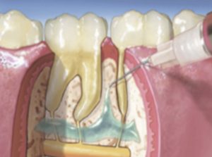
Most people find it a much more tolerable sensation than a traditional anaesthetic injection, it can be barely noticed. Occasionally it can be used as supplementary method to major oral surgery or for intense inflammation when traditional anaesthesia is not effective enough.
Digital implantation
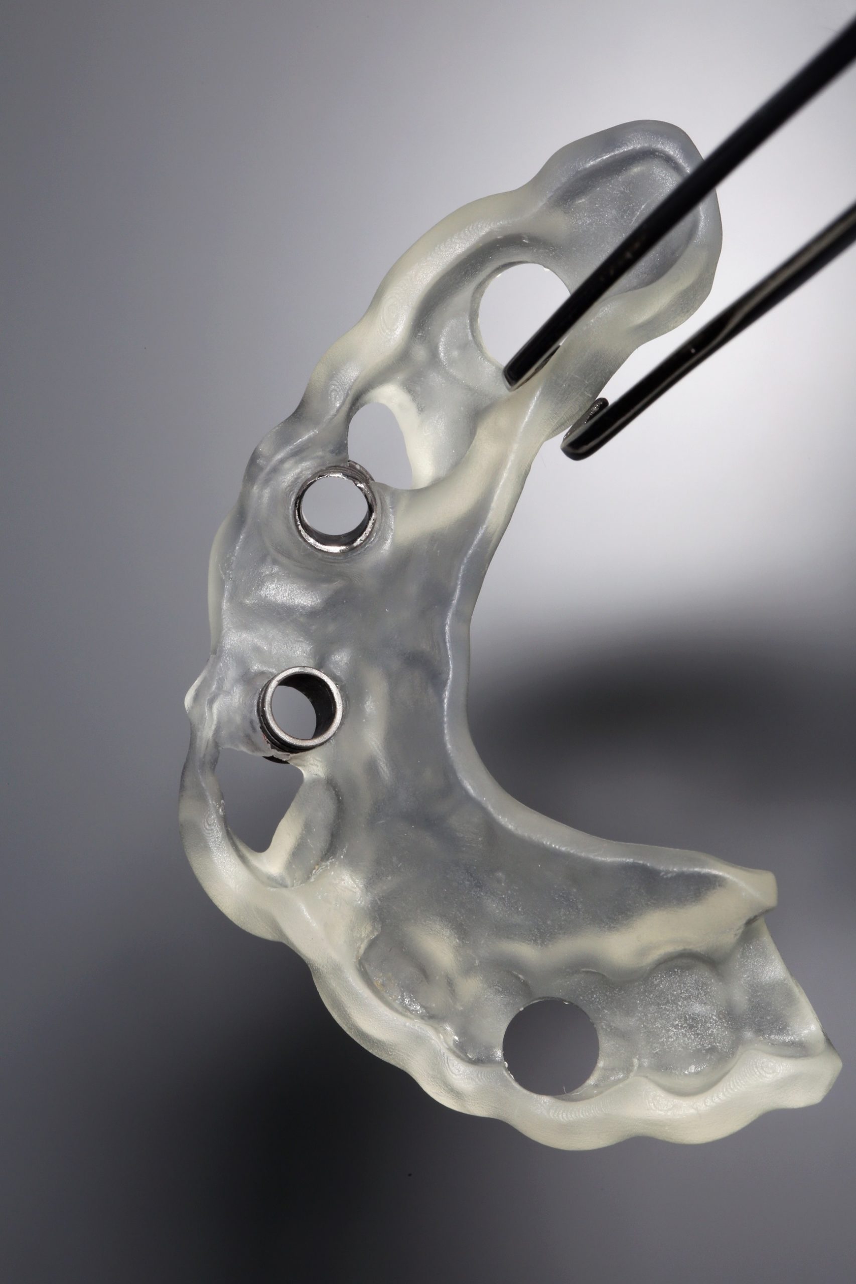
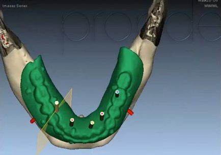
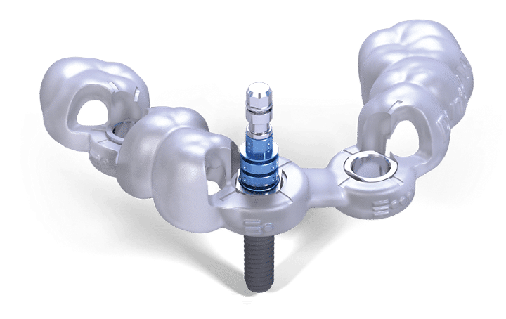
One of the most precise methods of implantation. It can solve many seemingly impossible cases where, for example, the proximity of a nerve would not allow hands-free surgery. After a digital CT scan, the surgery is planned on a computer. For the implant, which is digitally placed to an accuracy of tenths of a millimeter, a 3D printer prints a surgical tool that determines the direction and depth of the implant hole. The preparation of the hole and the implantation is then carried out through a gum wound of a few millimeters, to which the restoration will be subsequently fixed.
The process can be summarised as follows:
1. Medical consultation
Your dentist will inform you about the details of the treatment and the Surgical Guide.
2. 3 dimensional diagnostics
A CT scan of the area of tooth loss is taken to map the bone tissue and soft tissue. The scan is completely painless and takes only a few seconds.
3. Computer-aided surgical planning
Based on the CT scan, your dentist will plan the implantation procedure in a relaxed environment using a computer. Thanks to the planning software, the positioning and optimal size of the implants can be perfectly modelled, taking into account the amount and quality of bone available, as well as the location of the nerve canals and sinuses. The device shall be ordered and delivered to our practice within a few weeks.
4. Implantation procedure
A custom-made surgical guide – implantation template – based on the surgical plan is used to perform the procedure. The use of an implant template ensures that the implants are placed perfectly in their intended position and that you experience as little discomfort as possible.
5. Tooth replacement
Once the implants are placed in the optimal position, your dentist will prepare the most ideal prosthesis possible after the healing period.
